Oodinium (Velvet)
Eukaryota
Group Alveolates
Sub-group Dinoflagellata
Fish parasitic genera: Oodinium, Amyloodinium, Piscinoodinium, Credpidoodinium
Species include: P. limneticum, P. pillulare (O. pillularis), A. ocellatum
 Among the Dinoflagellates the best known are species which cause toxic blooms called "Red Tides." The organisms dealt with here are parasites of fish but as Dinoflagellates are taxonomically distinct from all other protists listed on these pages. While this group is often referred to as one of the algal groups it is sometimes misleading to apply the word algae to them. The nomenclature of these organisms has been revised recently and I will not attempt to discern among species or the genera except to note that Amyloodinium are the organisms encountered on fish in the marine environment and that there are other genera and species with non-fish hosts.
This parasite gained the name Velvet due to the appearance of the tan-colored coating it produces on heavily infected fish. It affects a wide range of fish species and can be deadly to fry and small fish. Infective spores attach to the skin of fish and develope penetrating rhizoids with which to feed off the host. The organism grows into a reproductive cyst which eventually falls off the fish and produces numerous spores while on the substrate. These dinospores become motile by a flagellum to seek out and infect a new host. In P. pillulare these posess a red eye-spot and sight is involved in locating the potential host. This needs to be accomplished within approximately 24 hours or the organism dies.
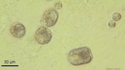
Oodinium cysts in a skin smear of a fish.
Diagnosis:
A presumptive diagnosis of this disease can be made on the basis of external signs. These include clamped fins, abnormal swimmimg, and a dusty look of the skin of the fish, these signs progressing through a population. Small fish may die soon after the onset of symptoms.
Microscopic observation of skin scrapings or fin samples should reveal large numbers of the parasitic stage. The cysts contain numerous spherical granuals of similar size, are greenish-brown in color, and are attached to the outer layer of skin on the fish. In some individual specimens a large clear-colored area in the center of the organism is discernable as the nucleus. The chitinous shell of the cyst is often visible as a thick cell wall as in the above photograph. The rhizoids are not normally visible but a side view of the organism may show signs of a colorless extension where attachment to the host is made. More than one parasite should be observed in order to form a confident diagnosis since no individual is likely to present all of the representative characteristics and because some contaminant organisms may resemble these parasites.
The motile stages are not commonly seen, although other dinoflagellates often find their way onto the microscope slide. In my opinion the presence or absence of motile dinospores, or the observation of motile dinoflagellates should not be considered in the diagnosis of the disease. Since sizes and shapes of the parasite cysts will vary I feel these characters should not weigh heavily in diagnosis either, though they may be of value in discriminating between species of parasite involved.
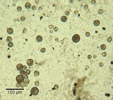
Numerous Velvet parasites on a Betta splendens
Treatment:
Acriflavin (Trypaflavin) is a commonly used and effective therapeutic for this disease and is suitable for most species of fish.
Copper compounds are effective and commonly used in marine aquaria. They should be used with care in freshwater aquaria and when dealing with fish having a low tolerance for copper.
Saltwater baths can be effective in treating freshwater species of the parasite and, conversely, freshwater baths are used in treating marine fish. Dipping freshwater fish in a strong salt solution for a fraction of a minute has been known to cause the parasites to fall off in some cases. I have found this method valuable in treating fish that were too heavily infested to survive a more lengthy treatment. Salt is sometimes added to the water (at one tablespoon to five gallons or stronger) to treat the disease or as a preventative. This may or may not be effective.
Quinine HCl is sometimes used in treatment of this disease but dosage needs to be figured accurately and maintained properly. A proprietary formulation should be used according to the instructions included.
Note; Although newly released dinospores of Oodinium need to find a host quickly in order to live there is some thought that the reproductive stage may be able to survive in the substrate for quite some time before producing spores under some conditions.