Tests could predict risk of dangerous heart rhythm after heart attack

Dec 17 (HeartCenterOnline) - A combination of tests could help physicians predict which heart attack survivors are at greater risk of experiencing life-threatening heart rhythms, according to a new study. Published in the current issue of the Journal of the American College of Cardiology, the study suggested that a combination of four or five tests could help classify up to 90% of heart attack survivors as "high risk" or "low risk" for life- threatening heart rhythms. Results of the study were based on an analysis of 44 published studies that included data from each of the tests. Based on their findings, the researchers proposed a "staged" approach, where most patients would only need to undergo four noninvasive tests, reserving the more invasive test for cases that remained unclear after the first four tests. The four noninvasive tests were the results of a signal- averaged electrocardiogram, the detection of any severe ventricular arrhythmias (irregular heart rhythms), the measurement of heart rate variability and the measurement of the left ventricular ejection fraction. The invasive test being investigated was the electrophysiology study. Each of these tests is briefly defined below. Like a standard electrocardiogram (EKG), a signal- averaged electrocardiogram (SAEKG) is a painless test that measures the heart's electrical activity. The difference is that an SAEKG uses a computer to strengthen certain electrical signals (late potentials), which most often occur in damaged and scarred portions of the heart muscle. Identifying these late potentials can be useful for evaluating a patient's risk of ventricular arrhythmias after a heart attack or heart surgery, identifying scar tissue from prior heart attacks and other functions. The left ventricular ejection fraction (LVEF) is the percentage of blood pumped out of the heart with each heartbeat. It is measured by either an echocardiogram or nuclear imaging, which provide images of the heart and reveal vital information about the heart's structure and function. Both heart rate variability and the presence of any severe ventricular arrhythmias were determined by an ambulatory EKG, which works just like a standard EKG except that people carry the small device with them rather than completing the test in a medical setting. The device monitors the heart rhythm either continuously or only when a patient is experiencing unusual cardiac symptoms, depending on which type of test is chosen. An electrophysiology study (EP study) is a procedure in which a thin tube (catheter) is inserted into a blood vessel (e.g., in the groin) and guided to the heart, where it can perform specific, essential measurements of the heart's electrical activity and pathways. An EP study can help assess the presence of arrhythmias or, in some cases, to stimulate the development of an arrhythmia in order to gauge the effectiveness of medical treatments. The study in the Journal of the American College of Cardiology was an analysis of past research, and the authors concluded that their promising findings should be applied to a prospective study. This means that heart attack survivors would be tested as proposed and then closely followed to see how well the tests were able to determine the "high risk" and "low risk" patients in the future. Patients identified as "high risk" might receive early treatment with antiarrhythmic drugs, an ICD or other treatment options.

electrophysiology Study
The Arrhythmia Center
The Cardiac Arrest Center
   
The ICD Center
The Heart Attack Center

Summary

Electrocardiogram An electrocardiogram (EKG or ECG) is a recording of the heart's electrical activity as a graph or series of wave lines on a moving strip of paper. This gives the physician important information about the heart. For example, it can show the heart’s rate and rhythm. It can also imply decreased blood flow (cardiac ischemia), enlargement (hypertrophy) of the heart or the presence of either current or past heart attacks.

EKGs are noninvasive, quick, safe, painless and inexpensive tests that are routinely done if a heart condition is suspected. Depending on what the results show, in combination with the patient’s medical history and a physical exam, the physician may order further tests or a combination of medications and lifestyle changes.

 
What is an electrocardiogram (EKG)?
An electrocardiogram (EKG or ECG) is a recording of the heart's electrical activity as a graph or series of wave lines on a moving strip of paper or video monitor. This allows the physician to evaluate the heart’s rate, rhythm and certain cardiac problems. As a result, the highly sensitive electrocardiograph machine can help the physician to detect various heart irregularities, disease and damage. For example, an EKG may be able to show whether a patient’s heart muscle was damaged as a result of a heart attack.
An EKG is routinely used when heart disease or damage is first suspected in a patient. For example, the patient may be experiencing symptoms such as the following:

The patient’s first EKG (a baseline EKG) will be compared with EKGs taken in the future, if they become necessary (e.g., after a heart attack).
 

What types of conditions might an EKG detect?
 
A physician can use EKGs in the diagnosis of a wide variety of abnormalities and diseases, such as the following: Coronary Artery Disease: Every 29 seconds, an American will experience a coronary event such as a heart attack, according to the American Heart Association.

Many people with coronary artery disease, heart valve disease or heart muscle disease will eventually have abnormal EKG readings. Because many EKGs are done while the patient is at rest, certain abnormalities that occur during periods of stress may not appear even in patients with significant disease. In fact, it has been estimated that the resting EKG is accurate only about 50 percent of the time. Because it is very common to see this false-negative result (i.e., the EKG doesn’t find the damage or abnormality that is really present), a normal EKG is not enough to rule out suspected heart disease.


How does a patient prepare for an EKG?
Patients should wear comfortable clothing that can be easily removed from the waist up. There is no need to limit eating or drinking before the EKG, and the test can be scheduled for any time of day. The test may be done in the physician's office, a hospital or a clinic.

How is an EKG done?

EKGs are noninvasive, quick, safe and painless. Once the patient has removed his or her clothes from the waist up, he or she will be instructed to lie down. A nurse or technician will thoroughly clean a total of ten areas on the patient’s chest, arms and legs. The technician will apply gel to these areas. Ten small metal devices (electrodes) are then attached to those clean areas of the skin and connected by wires to the electrocardiograph machine. The technician will enter the following information about the patient into the computer:
  • Name
  • Age
  • Sex
  • Date

Once this is done, the patient will need to lie perfectly still for about one minute while resting-heart activity is measured and recorded. Each electrode produces a "tracing" or “lead” of a particular area of the heart and its activity. As a stand-alone test, the entire EKG takes about 5 to 10 minutes.

There may be a portion of the evaluation that includes a form of exercise stress test. This test requires the patient to either walk on a treadmill at varying speeds and elevations, or ride a stationary bicycle, to measure the heart’s activity during physical activity.
 

What happens after the EKG?
 
EKG results should be immediate. In some cases, they can be done outside the physician’s office and transmitted via phone line back to either the physician or another diagnostic center for evaluation. Transtelephonic monitoring, for the purpose of assessing the heart rhythm, can detect irregular heart rhythms (arrhythmias) that occur when the patient is not in the physician’s office.

Echocardiogram Depending upon the results, further treatment may or may not be necessary. If damage or a problem is found, usually a combination of medications and risk-reducing lifestyle changes is prescribed. Also, additional tests are usually ordered. Based on the results of the non-stress EKG, an
exercise stress test or a nuclear stress test may be done inconjunction with an echocardiogram. An echocardiogram uses sound waves to track the structure and function of the heart. If abnormalities are not detected at the physician’s office, then the patient may be asked to wear a Holter monitor, a portable tape recorder that records the heart’s rhythm, usually over a 24-hour period.
The P wave, the QRS complex and the T wave

 
The heart’s electrical system is quite complex.  Conduction System: The heart has its own electric pacemaker, which regulates the heartbeat. Specialized nerves send signals to the pumping chambers, telling them to contract. Electrical rhythms begin as impulses emitted from the sinoatrial node, also known as the heart’s “natural pacemaker.” The impulse then travels across a specific route, or pathway, traveling through the AV node and into the ventricles. Once they reach the ventricles, the impulses serve as a set of instructions, causing the chambers to contract in a routine and consistent manner. If rhythms are interrupted, delayed or sent down the wrong path, the heartbeat may become irregular, too fast or too slow. These rhythms are detected by an EKG.

The EKG breaks down each heartbeat into a series of electrical waves that give important information about the heart. Three distinct waves are displayed: the P wave, the QRS complex and the T wave. The P wave is associated with activity in the heart’s upper chambers (
atria). The QRS complex and the T wave both reflect activity in the heart’s lower chambers (ventricles). These three waves are critical in diagnosing a wide variety of heart-related conditions.
 

Summary

Electrocardiogram Similar to the standard electrocardiogram (EKG), the signal-averaged electrocardiogram (SAEKG) is a painless test used to assess whether a patient is at high risk of developing potentially fatal heart rhythms such as ventricular tachycardias. After changing into a hospital gown from the waist up, the patient will need to lie still for a few minutes while this test is being performed. Afterward, the patient can get dressed and return to his or her usual daily activities immediately. If an irregular heart rhythm (arrhythmia) is suspected, then the physician may order a more invasive test called an electrophysiology study.


What is a signal averaged EKG?
A signal-averaged electrocardiogram (SAEKG) is a painless test that is very similar to the standard electrocardiogram (EKG), which measures electrical activity of the heart. However, the SAEKG uses a computer to strengthen certain electrical signals (late potentials) and reduce the level of random noise that surrounds them. The EKG signals are filtered and parts of them are averaged, which allows identification of the late potentials and provides physicians with information about whether the patient is at risk for developing potentially lethal heart rhythms.

Like a standard EKG, the SAEKG displays the heart’s electrical activity as three distinct waves: the P wave, the QRS complex and the T wave. The P wave is associated with activity in the upper chambers of the heart (atria), and the QRS complex and T waves reflect the activity in the lower chambers of the heart (ventricles). After a computer has filtered these waves, the test can reveal the presence of any late potentials, which most often occur in damaged and scarred portions of the heart muscle. Identifying these late potentials can be useful for:
  • Evaluating a patient’s risk of ventricular arrhythmias after a heart attack or heart surgery
  • Diagnosing the cause of unexplained fainting (syncope)
  • Detecting heart chamber abnormalities
  • Identifying scar tissue from prior heart attacks

Tachycardia: An unusually fast heartbeat (more than 100 beats per minute). It may be a sign of an arrhythmia, or it may be the result of normal factors (e.g., fever or exercise). An SAEKG is rarely useful if performed immediately after a heart attack, because it may take several hours for any scarring to occur. Late potentials usually occur within the first week after the heart attack. About 70 to 90 percent of patients with ventricular tachycardias demonstrate late potentials on the SAEKG.


Also known simply as an "echo," an echocardiogram of the heart's chambers and valves is called a transthoracic echocardiogram. The word "transthoracic" means "across the chest". It is a painless test that is very similar to an x-ray but without the radiation. Instead of using x-rays, it uses high-frequency sound waves (ultrasound) to get a picture of the four heart chambers and the four heart valves. Echocardiogram The sound waves bounce back from the heart chambers and valves, producing images and sounds that can be used by the physician to detect damage and disease. Because it does not involve any of the radiation that an x-ray does, it is a very safe test. In fact, it uses the same technology that is used to evaluate a baby's health before birth.

To do a transthoracic echocardiogram, the physician or technician prepares the chest area by applying a conductive gel. A small device called a
transducer is then placed on the patient's chest, and a picture of the area is seen immediately on a video monitor. At that time, the lights in the room may be dimmed to give a better view of the various monitors that are recording the results of the echocardiogram.

There are different types of echocardiograms, which include the following:
  • One-dimensional (M-mode; motion mode). A one-dimensional view of the heart, as if a line were drawn straight through it.

     
  • Two-dimensional (cross-sectional). A two-dimensional view of the heart, which shows both length and width of heart structures.

     
  • Doppler ultrasound. A test that measures the speed with which blood is traveling through the heart. Although echocardiograms are generally black and white, a color Doppler adds color for better viewing of the blood flow patterns.

     
  • Stress echocardiogram. An echocardiogram that is performed while the patient exercises in a controlled manner on a treadmill or stationary bicycle at varied speeds and elevations. The wall motion of the heart's pumping chamber before and immediately after exercise may reveal a lack of blood supply to selected areas of the heart muscle.

     
  • Chemical (e.g., dobutamine or adenosine) stress echocardiogram. A type of stress test that is used with patients who are unable to perform physical activity. A chemical stress echocardiogram measures the reaction of the heart under chemically induced stress, in order to assess the wall motion of the heart muscle. A drug such as dobutamine causes the heart to react as if the person were exercising, though the patient is actually at rest.

     
  • Transesophageal echocardiogram (TEE). A minimally invasive echocardiogram that requires a transducer to be inserted down the patient's throat into the esophagus (the long tube that connects the throat to the stomach). Because the esophagus is located so close to the heart, very clear images of the heart structures and valves can be obtained from the inside of the body, without the interference of the chest wall and lungs.

There is also a type of echocardiogram called the intravascular echocardiogram, in which a transducer is threaded into a blood vessel via a catheter and provides information about plaque and calcium deposits on the inside of the blood vessel.


What is an echocardiogram?
 
Depending on which type of echocardiogram is used, an echocardiogram is either a noninvasive or minimally invasive diagnostic test used to measure the structure and function of the heart. By obtaining this information, the physician can:
  • Check the health and performance of heart valves
  • Measure the heart wall and check for abnormalities in heart wall motion
  • Detect disease or accumulation of fluid in the pericardium
  • Identify blood clots

An echocardiogram uses a hand-held device called a transducer to send and receive ultrasound (high frequency sound waves) signals. A computer then calculates the travel time of these sound waves to and from the heart and constructs an image of the heart onto video monitors, paper, videotape and/or digital storage devices.

An echocardiogram does not require the use of radiation and produces a very reliable image, making it an exceptionally safe and painless test. As this is the same technology used to evaluate a baby's health before birth, it also considered a safe method for examining children and pregnant women.

An echocardiogram is often given to patients who have had experienced signs and symptoms such as the following:

What are the types of echocardiograms?
 
There are many different types of echocardiograms, which include the following:
  • One-dimensional (M-mode; motion mode). A one-dimensional view of the heart, as if a line were drawn straight through it.

     
  • Two-dimensional (cross-sectional). A two-dimensional view of the heart, which shows both length and width of heart structures.

     
  • Doppler ultrasound. A test that measures the speed with which blood is traveling through the heart. Although echocardiograms are generally black and white, a color Doppler adds color for better viewing of the blood flow patterns.

     
  • Stress echocardiogram. An echocardiogram that is performed while the patient exercises in a controlled manner on a treadmill or stationary bicycle at varied speeds and elevations. The wall motion of the heart's pumping chamber before and immediately after exercise may reveal a lack of blood supply to selected areas of the heart muscle.

     
  • Chemical (e.g., dobutamine or adenosine) stress echocardiogram. A type of stress test that is used with patients who are unable to perform physical activity. A chemical stress echocardiogram measures the reaction of the heart under chemically induced stress, in order to assess the wall motion of the heart muscle. A drug such as dobutamine causes the heart to react as if the person were exercising, though the patient is actually at rest.

     
  • Transesophageal echocardiogram (TEE). A minimally invasive echocardiogram that requires a transducer to be inserted down the patient's throat into the esophagus (the long tube that connects the throat with the stomach). Because the esophagus is located so close to the heart, very clear images of the heart structures and valves can be obtained from the inside of the body instead of the outside.

Each type of echocardiogram provides unique information to the physician regarding the heart structures and function. For example, a one-dimensional echocardiogram is very helpful in determining the size of one of the heart's chambers (atria and ventricles), thickness of the chamber walls, function of the mitral valve and weight of the left ventricle. The two-dimensional echocardiogram is particularly helpful in seeing the relation of the heart's chambers to each other, the ejection fraction (a measurement of how much blood is pumped from the heart), and problems with the heart's valves. Doppler ultrasound and its colorized version (the color Doppler) are helpful in detecting a variety of problems, including those of the valves or of the septum (the muscular wall separating the left and right ventricles).

These three types of echocardiograms (M-mode, two-dimensional and Doppler) are particularly useful for diagnosing the following conditions:

  • Valvular heart disease. Types of this disease include a condition in which the valves have narrowed (valvular stenosis: mitral stenosis, aortic stenosis, tricuspid stenosis, or pulmonic stenosis), and a condition in which the valves are leaking (valvular regurgitation mitral regurgitation, aortic regurgitation, tricuspid regurgitation or pulmonic regurgitation).

     
  • Rheumatic heart disease. The effects of rheumatic fever that contribute to major problems with the heart's valves, chambers and vessels.

     
  • Bacterial endocarditis. An infection in one or more of the heart valves.

     
  • Cardiomyopathy. A disease in which the heart muscle is unusually thick, stiff, dilated or weak.

     
  • Congestive heart failure. A condition in which blood flow and circulation are not adequately maintained by one or more valves or chambers of the heart.

     
  • Pericarditis. Inflammation of the pericardium (a thin, fluid-filled sac surrounding the heart).

     
  • Tumors in the heart.

     
  • Coronary artery disease. The obstruction of blood flow to the heart and the body due to hardened arteries (atherosclerosis).

     
  • Cardiac ischemia. A condition in which the heart is not getting enough oxygen, usually because blood flow is restricted by hardened arteries (atherosclerosis).

     
  • Heart attack. Scarring, or death, of heart muscle due to oxygen deprivation from a closed artery.

     
  • Shunt. Abnormal connection between the heart's chambers.

The physician may also do a stress echocardiogram or “stress echo” to see how the heart functions during physical activity. Usually, a stress echocardiogram involves doing the echocardiogram while the patient is exercising on either a treadmill or a stationary bicycle, at varying speeds and elevations. However, if the patient is unable to perform this physical activity, the physician may choose instead to do a chemical (e.g., dobutamine or adenosine) stress echocardiogram. This is a test in which the patient is given a medication that causes the heart to beat more strongly, showing the physician what the heart would be doing if it were beating under the more standard stress of exercise.

In some cases, none of these echocardiograms can provide the physician with enough information. For example, the tests may not give accurate information if the patient has been diagnosed with either obesity (weighing more than 20 percent of his or her ideal weight) or chronic obstructive pulmonary disease (such as emphysema or chronic bronchitis). Alternatively, the physician may be concerned that a blood clot is present in one of the heart's upper chambers (atria), or that fatty plaque is present in the aorta, which are located deep in the chest. In these situations, the physician may choose to do a more invasive form of the test called a transesophageal echocardiogram (TEE), which gives a very clear view of the heart from the inside of the body instead of the outside.

How can patients prepare for echocardiograms?
Other than wearing loose-fitting clothing, there are no special restrictions or preparation prior to having most echocardiograms done. In general, patients may continue to eat, drink fluids and take prescribed medication as normal. However, people who are scheduled for either a transesophageal echocardiogram (TEE) or a chemical stress echocardiogram are often told to stop eating and drinking for about four to six hours before the test. This helps to prevent any nausea or vomiting during the test. Also, people scheduled for a TEE may be asked to reduce or stop certain medications that they usually take.

How is a noninvasive echocardiogram done?

An echocardiogram is performed in a variety of settings, including hospitals, cardiac labs, testing centers or the physician’s office. Most noninvasive echocardiograms take about 30 minutes. The chemical stress echocardiogram takes 1 to 1.5 hours. Additional time may also be needed for the physician or technician to record information about the patient and to answer all of the patient’s questions. People scheduled for any of the noninvasive echocardiograms are free to drive after the test.
Patients are required to remove clothing from the waist up, and are given a hospital gown to wear during the test. The physician or technician will prepare the chest area by applying a conductive gel. People scheduled for a chemical stress echocardiogram will receive an intravenous (I.V.) line via a needle inserted into a vein at the back of the wrist. The I.V. line allows medications to be given to the patient without having to stick the patient with more needles.
A hand-held device called a transducer is then placed on the chest directly over the gel and pictures of the chest are seen immediately. At that time, the lights in the examination room may be dimmed to allow a better view of the various monitors that are recording the results of the echocardiogram.
An electrocardiogram (EKG) may also be performed during this test. This is a painless test in which about 10 to 12 small metal devices (electrodes) are taped to thoroughly cleansed areas of the chest and attached by wires to the electrocardiograph machine. The EKG gives information about the electrical activity of the heart and can help detect irregular heart rhythms (arrhythmias).
Throughout the echocardiogram, the physician or technician may change the position of the transducer, reposition the patient or ask the patient to take certain actions, such as holding his or her breath. Each of these changes helps to create clearer images of the heart.
Following the echocardiogram taken while the person is at rest, people scheduled for a chemical stress echocardiogram will also have an echocardiogram taken after a medication such as dobutamine has been given through the I.V.
When the test is complete, the transducer is removed and the gel is cleansed completely from the chest area.
What happens after an echocardiogram?
There should be no side effects or complications from an echocardiogram, although people who had a chemical stress echocardiogram may sometimes experience some minor side effects from the medication such as:
  • Nausea (upset stomach)
  • Palpitations (strong, fast or obviously irregular heartbeat)
  • Numbness in the arms or legs
  • Flushing or a wave of warmth
  • Chest pain
  • Headaches

The medication may also cause complications due to stressing the heart, which include irregular heart rhythms (arrhythmias) or low blood pressure hypotension.
Occasionally a child undergoing a pediatric echocardiogram will be given a calming medication (sedative). The patient’s parent or guardian should discuss with the physician the potential effects of a sedative both during and after the examination.
 

What is a transesophageal echocardiogram?
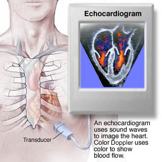
During a transesophageal echocardiogram (TEE), a small transducer attached to a tube (echoprobe) is inserted into the esophagus via the mouth and throat. This will not affect the patient’s ability to breathe freely but may temporarily interfere with swallowing. Once positioned in the esophagus, the transducer can transmit very clear images of the heart’s size, structures and functioning.

A TEE takes up to 90 minutes. Additional time may also be needed for the physician or technician to record information about the patient and to answer all of the patient’s questions. People who are scheduled for a TEE are encouraged to arrange for transportation from the test, because they will be given a calming medication (sedative) that often leaves them feeling groggy or light-headed.

Just before the TEE, the patient will be asked to remove dentures and oral prostheses and to lie down on his or her left side on the examination table. An intravenous (I.V.) solution is started and a mild calming medication (sedative) can be administered. Heart and blood pressure monitoring will begin, and will continue throughout the procedure. Finally, an anesthetic spray is sprayed into the throat to reduce the gag reflex.
The physician will insert the echoprobe and feed it to the esophagus, just behind the heart. The patient may be asked to swallow in order to help move the echoprobe into location. Some discomfort at this stage is normal. Once in place, imaging begins. The transducer at the tip of the probe may be periodically repositioned or advanced.
When imaging is completed, the echoprobe is withdrawn. Monitoring of vital signs will continue after the procedure until the sedative wears off. At that time, the patient will be able to leave the hospital or diagnostic center. People cannot eat or drink anything until the anesthetic spray has worn off and the gag reflex is restored, or else they could choke. This takes approximately one hour after the procedure.
Complications are rare, but could include:

People who have additional questions are encouraged to speak with their physician.