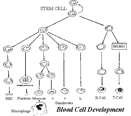
WHOLE BLOOD is
comprised of plasma (the liquid part) and the formed elements (red
blood cells, white blood cells, and
platelets).

The process by which all formed elements of the blood
are produced (hematopoiesis), occurs mostly in the bone marrow, where cells
mature from a primitive stem cell.
Billions
of red, white blood cells and platelets are produced per kilogram of body
weight daily. Factors important
in regulating blood cell production include the environment of the bone
marrow, interactions among cells, and secreted chemicals called growth
factors.
Patients with Waldenstrom’s Macroglobulinemia
experience a reduced capacity to produce several types of
whole blood cells in the bone marrow (myelosuppression)
because the overproduction of immature
WM cells suppresses production of the other blood cell types.
Chemotherapeutic agents (which destroy fast growing cells of the body)
also contribute to lowered blood cell production.
NOTE: As used in this paper, “Normal” is not what you might
presume. Nobody really knows what “normal” means, and it varies with
gender, age, nutritional environment and methods of testing. Laboratories
compare your readings to a “Normal” or “Reference Range” but that
range is not a national standard,
but a comparison with all of their other tests. We collected “ranges” from
18 major national clinics and labs, and all of the numbers differed. The
numbers you see here will give you some rough idea of “normal” but what is
more important is how your numbers change over time, more than the absolute
value. It is for this reason that it is important to always have your blood
tests done at the same laboratory.
A
COMPLETE BLOOD COUNT (CBC)
is a measurement of the blood cells in a specific volume of blood. Common
components of a blood count that are important to WM are as follows:
RBC
Name:
Red Blood Cells
(erythrocytes).
Normal Range: 4.7-6.1 million/cu.mm
WM Abnormality: Low RBC count (anemia) diminishes the body’s ability to carry oxygen
to the tissues.
Function/Test Purpose: Formed
in the bone marrow, a typical red blood cell lives for about 120 days. These
cells transport oxygen to all parts of the body. This test is done to support
other tests in diagnosis of anemia and to supply figures for computing the
erythrocyte indices, which reveal RBC size and hemoglobin content.
Test Mechanism: Red Blood Cells are
counted with an instrument called a Coulter Counter. The sample is diluted in
an electrically charged solution and moves slowly through an aperture across
which a specific voltage passes. As each cell passes through, the voltage
changes, creating a pulse. The voltage magnitude varies also with cell size.
This way the cells are counted, and sized.
Particles greater than 36 fL are counted as RBCs.
HCT -
Name:
Hematocrit
Normal
Range: 42-51%
WM
Abnormality:
Lowered hematocrit.
Function/Test Purpose: The volume occupied by the packed red blood cells
in a given volume of centrifuged blood. It
is used in determination of anemias, and is usually expressed as a percentage of the volume of the
whole blood sample.
Test Mechanism: This is a calculated value from the values of the red cell count and
the Mean Corpuscular Volume.
HCT = RBC x MCV
HGB
Name:
Hemoglobin
Normal
Range: 14-18 g/100 ml.
WM
Abnormality: Lowered
hemoglobin.
Test Purpose: Hemoglobin
is the pigment in red blood cells that contains iron and transports oxygen to
the tissues. This is the main component of the red blood cell. The test coordinates
with other red blood cell data.
Test Mechanism: An instrumental method, using a spectrophotometer, measures the
intensity of light which passes through the blood sample. Less light
transmittance equates to more hemoglobin.
----------------------------------------------------------------------------------------------
ERYTHROCYTE INDICES below provide information about the size, hemoglobin
concentration and hemoglobin weight of an average RBC:
MCV
Name: Mean Corpuscular
Volume
Normal
Range: 80-96 fL
WM
Abnormality: Varies;
however, MCV may be elevated due to erythrocyte aggregation.
Function/ Test
Purpose:
This is the average volume of the red blood cells, the ratio of hematocrit to the RBC count. The
Mean Corpuscular Volume expresses the average size of the erythrocytes, and
indicates whether they are undersized, oversized or normal.
Test Mechanism: Measured with a Coulter Counter. The cell volume is derived
from the amount of voltage variance as each cell is counted.
MCV = HCT
x 10
RBC count (millions/μl)
MCH
Name:
Mean Corpuscular Hemoglobin
Normal
Range:
20-33 pg
WM
Abnormality: Varies;
hemoglobin will be depressed if RBC count is lowered.
Function/Test Purpose: Mean
Corpuscular Hemoglobin is the content of hemoblobin in the average red blood
cell, the hemoglobin/RBC ratio. It
expresses the weight of
hemoblobin in an average RBC.
Test
Mechanism: A calculated value using RBC count, hemoglobin and
hematocrit.
MCH = Hemoglobin (g/L) x 10
RBC count (millions/μl)
MCHC
Name: Mean Corpuscular
Hemoglobin Concentration
Normal
Range: 32-36%
WM
Abnormality:
Varies; if hemoglobin is lowered, hematocrit is also lowered.
Function/Test
Purpose: The ratio of hemoglobin weight to hematocrit.
This test defines the concentration of hemoglobin in
100 ml. of packed red blood cells. It helps distinguish the normally colored
cells from paler cells to help classify different anemias and aid in
determining cause.
Test
Mechanism:
A calculated volume using RBC count, hemoglobin and hematocrit.
MCHC = Hemoglobin (g/dl) x 100
HCT (%)
ESR
Name:
Erythrocyte Sedimentation
Rate
Normal
Range: 0-30 mm/hr in people over 50
WM Abnormality: Elevated rates are usually
found in people with WM because of the presence of excessive amounts of the
macroglobulin IgM.
Function/Test Purpose: This
is a determination of erythrocyte and/or plasma abnormalities.
It aids in the ability to distinguish among diseases with similar
symptoms.
Test Mechanism: This test establishes the
distance that erythrocytes have fallen in a vertical column of anticoagulated
blood under the influence of gravity. The
test procedure itself is easily influenced by a number of environmental
factors, including blood cell size and shape, temperature, handling.
This test does not stand alone as
a good diagnostic tool.
PLT
Name: Platelets (
thrombocytes)
Normal Range: 150,000 - 500,000/cu.mm
WM Abnormality: Low platelet count (thrombocytopenia)
and function. Platelets become coated with IgM molecules, decreasing their
clotting capacity.
Function/Test Purpose: Some
of the same stem cells that produce the red blood cells in the bone marrow
develop into large specialized
cells called Megakaryocytes; each of these is packed full of as many as 4000
platelets (discs with a sticky surface substance); the large cells
erupt and the separate platelets circulate throughout the bloodstream
to assist in clotting, surviving for about ten days.
Test Mechanism: During
the Coulter Counting cycle, as the platelets and RBCs pass through the
apertures, the particles which are between 2 & 20 fL in size are counted
as platelets.
MPV
Name:
Mean Platelet Volume
Normal
Range: 7.2-11.1 %
WM
Abnormality: Varies;
if platelets are depressed, platelet hematocrit will be lowered.
Function/Test Purpose: The
ratio of Platelet Hematocrit to Platelet Count; aids in determining platelet
volume and size.
Test
mechanism:
This is a calculated value. MPV=
Platelet Hematocrit
Platelet
Count
-----------------------------------------------------------------------------------------
WBC
Name: White Blood Cells
(Leukocytes) are composed of Basophils, Eosinophils and
Neutrophils, which are collectively called Granulocytes.
Other white blood cells are Lymphocytes
and Monocytes.
Normal
Range: 4.8-10.8 thousand/cu.mm
WM
Abnormality: Lowered WBC
count (leukopenia)
Test Purpose: Primarily formed in the bone marrow; white blood cells may also be
produced in organs of the lymphatic system such as spleen, thymus and lymph
nodes. They serve to fight off infections and protect the body from disease.
This test serves to monitor
disease progression and/or response to chemotherapy.
Test Mechanism: White Blood
Cells are counted with an instrument called a Coulter Counter. The sample is
diluted in an electrically charged solution which is passed slowly through an
aperture across which a specific voltage passes. As each cell passes through,
the voltage changes, creating a pulse. The voltage magnitude varies also with
cell size. This way the cells are counted, and sized.
--------------------------------------------------------------------------------------
WHITE
BLOOD CELL DIFFERENTIAL:
The
following percentages are part of the White Blood Cell Diffferential, and are
part of the Complete Blood Count.. They
evaluate the distribution of the various types of white blood cells based on
size distribution as determined by the Coulter Counter.
The sizes are then graphed to show numbers of each type. They are
compared to the normal range for a healthy person and abnormalities are
flagged. The
differential is done to evaluate the body’s capacity to resist and overcome
infection.
%
NEUT
Name: Percent Neutrophils
Normal Range: 7.4-10.4 thousand/cu.mm (50-70%
of WBC’s)
WM Abnormality: Lowered count and percentage, from disease or chemotherapy. However, if
other white blood cell counts also fall, percentage may not vary.
Function/Test Purpose: Neutrophils
are the most numerous white blood
cells; they are mobile cells that capture
foreign particles and bacteria that have entered the body, engulf and digest
them . These cells are often
referred to as Phagocytes.
The test is performed to relate Neutrophil count to other blood counts.
Test Mechanism: These are among the Granulocytes which are identified by Coulter Counter at 160-450 fL. They are
then differentiated from other Granulocytes by light scatter based on cell
shape and plotted.
%
LYMPH
Name: Percent Lymphocytes
Normal
Range: 20-30% of white blood cells
WM Abnormality: Change will depend
on status of disease and chemotherapy. Direct counts may fall; percentage may
remain the same. Most patients do not have large numbers of circulating WM
cells in the blood.
Function/Test Purpose: Originating
as immature stem cells in the bone marrow, they mature in the lymphatic
tissues to B cells (20% of the lymphocytes)
which release antibodies (Immunoglobulins
IgD,IgE,IgA,IgG,IgM) for defense against infectious agents, and to T cells
(80% of the lymphocytes), which provide cell mediated immunity.
As the lymphocyte matures it develops antigens (specific proteins) on
its own cell membrane. These are referred to as markers (in WM, the CD20
marker is identified) which
stimulate the production of the immunoglobulins. Once the cell fully matures,
and is referred to as a plasma cell, it usually no longer exhibits these
antigens. It is these antigens
which stimulate the excess production of IgM in the rapidly multiplying B
Lymphocytes of WM patients.
Lymphocytes are
responsible for specific recognition and immune responses to viruses, cancer
cells, and other foreign substances within the body.
The antibodies produced by the B cells of a healthy person coat the
foreign antigens, marking them for attack by the Neutrophils.
In WM, the excess antibody doesn’t find enough
foreign antigens to coat, so it tends to coat the blood platelets,
inactivating them, and it deposits in body tissues.
Both B and T cells are long lived, (about four years) and travel
throughout the body.
The test is performed to relate count to normal, and
to monitor disease progression.
Test Mechanism: Are nucleated
cells of 35-90 fL as measured by Coulter Counter.
%
MONO
Name: Percent Monocytes
Normal Range:
1.7-9% of WBC’s
WM Abnormality: Direct count may fall from disease or chemotherapy; if other white
blood cell counts fall, percentage may not be affected.
Function/Test Purpose: Monocytes
are a type of white blood cell produced in bone marrow, arising from the same
stem cell as the Neutrophil. They are mobile in the bloodstream or in body
tissues where they mature to cells called Macrophages.
Monocytes capture and destroy bacteria and other foreign substances,
remove dead cells from the body, participate in iron metabolism, and process
information about foreign antigens for the lymphocytes.
The test is performed to relate count to normal, and to monitor disease
progression.
Test Mechanism: 90-160
fL as measured by Coulter Counter.
%
EOSIN
Name: Percent Eosinophils
Normal
Range: 0-7% of WBCs
WM Abnormality: Direct count may fall from
disease or chemotherapy; if other white blood cell counts fall, percentage may
not be affected.
Test Purpose: Eosinophils
are a type of white blood cell that engulfs antibody labeled substances and
functions in inflammatory reactions. The
test is performed to relate count to normal, and to monitor disease
progression.
Test Mechanism: These
are among the Granulocytes which are
identified by Coulter Counter at
160-450 fL. They are then differentiated from other Granulocytes by light
scatter based on cell shape and plotted.
%
BASO
Name: Percent
Basophils
Normal
Range: Less than 1% of WBCs
WM Abnormality: Direct count may fall from
disease or chemotherapy; if other white blood cell counts fall, percentage may
not be affected.
Test Purpose: Basophils
are a type of white blood cell which secrete chemicals to promote inflammatory
reaction in the body. The test is
performed to relate count to normal, and to monitor disease progression.
Test Mechanism: These are among the
Granulocytes which are identified
by Coulter Counter at 160-450 fL. They do not show up by light scatter, but
demonstrate a different conductivity from other Granulocytes and are that way
differentiated.
-----------------------------------------------------------------------------------------
ABS
NEUT
Name:
Absolute Neutrophil Count
Normal
Range: 7.4-10.4 thousand/cu.mm
WM Abnormality: Lowered Neutrophil count (Neutropenia);
puts the patient at greater risk from infectious agents both inside and outside
the body.
Test Purpose: Neutrophils
are a type of mobile white blood cells that capture foreign particles and
bacteria that have entered the body, engulf and digest them ; these cells are
often referred to as phagocytes. The
test is performed to identify abnormal Neutrophil count and relate to disease
progression.
Test
Mechanism:
This is a calculated value:
Absolute Neutrophil Count = Total white blood cell
count x % neutrophils
100
VISCOSITY
Name:
Serum Viscosity Test
Normal
Range: 1.0-1.8
WM
Abnormality: Increased blood viscosity (hyperviscosity)
Function/Test Purpose: Viscosity
is the property of blood that resists a force causing it to flow. It is compared
to the viscosity of distilled water at room temperature, and is a function of
the concentration of protein in the blood. High serum viscosity is most often
caused by an overabundance of IgM.
Test Mechanism: Blood is allowed to
clot. Then the serum is separated by centrifuging and warmed. The serum is
allowed to flow down through a narrow tube (viscometer), and its rate of flow is
timed. It is compared to the rate of flow for distilled water.
Serum Viscosity =
Flow time of blood serum
Flow time of distilled water
IMMUNOGLOBULINS
Blood
serum total proteins are grossly separated into Albumin and Globulins.
Albumin
is the protein of highest concentration in the serum and is important for
maintaining oncotic blood pressure (keeps the blood from leaking out into the
tissues).
Globulins
are divided into alpha, beta, and gamma globulins, based on the spacing zones
into which they are graphed when separated, and include the types D, E, G, A
& M (called immunoglobulins). Those
of interest in WM are the immunoglobulins A, G and M.
Name:
Immunoglobulins A, G, & M
Normal
Range:
Immunoglobulin A:160-260 mg/dl
Immunoglobulin G: 950-1550 mg/dl
Immunoglobulin M:
50-300 mg/dl. This molecule is approximately six times larger than the
others.
WM Abnormality: IgM count is extremely
elevated; can be up as much as 10,000 mg/dl, and is the major blood abnormality
which leads to diagnosis of this disease. IgA
and IgG count may be depressed.
Function/Test Purpose: Immunoglobulins are found in the blood serum; they are
antibody proteins produced by the B white blood cells at an immature stage as
part of the immune function to mark foreign antigens for destruction.
The test is performed to determine globulin count and to monitor disease
progression.
Test Mechanism: The
laboratory test to separate and quantify the globulins is called Serum Protein
Electrophoresis. This is an analysis of the proteins present in blood serum
based on size and electrical charge. The blood serum (fluid portion of blood) is
placed on specially treated cellulose acetate paper, saturated in an
electrostatic fluid, and exposed to
a continuous electric current. The various proteins migrate (move on the paper)
to form bands that indicate the relative proportion of each protein fraction.
The proteins are then stained and a densitometer can determine and graph
the amount of each protein present by the intensity of stain.

BONE
MARROW BIOPSY
WM
Abnormality: Marrow shows a proliferation of immature B Lymphocytes
and depression of other cell
types. The
result is reported as “% involvement of nucleated marrow cells” and
diagnosis is confirmed at greater than 30%.
Function/Test Purpose: A
bone marrow biopsy is an examination of the soft tissue within the bones (bone
marrow); it provides reliable diagnostic information about blood disorders.
It is used to diagnose anemias, thrombocytopenia, to evaluate the
effectivenes of chemotherapy and help monitor suppression of blood cells of the
bone marrow . In WM it is
also used to identify infiltration of bone marrow by the WM cells.
Test Mechanism: A local anaesthetic is
administered, and sometimes a sedative. Patient will feel pressure on needle
insertion, and a brief pulling pain on removal of the marrow. Marrow specimen is
placed on a series of slides and sent to the laboratory for microscopic
examination. Special stains are
used to obtain count of different types of marrow cells for identification of
disease cells.
CD20
MARKER
Name:
Cell Marker Identification
WM
Abnormality: WM disease cells may exhibit CD20 marker
Function/Test Purpose: This test aids in diagnosis and monitoring
accumulation of WM cells at specific sites.
It is performed by a technique
called Flow Cytometry which is used for defining and enumerating lymphocytes. It
can detect, count and identify characteristic molecules on groups of cells.
Test Mechanism: Individual cells are
first tagged with fluorescing antibody
chemicals. These
chemicals will bind to a specific cell surface protein, a marker which
identifies that cell. Once the
antibody-marker complex is formed, the cells are now considered labeled.
The sample is then forced through a nozzle creating a fine stream of liquid
containing cells spaced singly at intervals.
The stream of cells then passes through a laser beam. They scatter the
laser light and the dyes on the labeled cells fluoresce. Phototubes pick up this
light. This provides information on the size and characteristics of the cell and
on the binding of the antibodies, expressing the surface markers.
Cells can then be separated by a cell sorter: as each cell passes
through the nozzle the light sends a
signal to the computer which
generates an electric charge characteristic of that particular cell. The charges
can be deflected and grouped - a specific charge representing each of the cells
with the same antibody-marker
complex. Thus an entire grouping of
cells expressing the CD20 marker has been identified and tracked.
UNIT LABEL CODE
g = gram
mg = milligram (one thousandth of a gram)
pg = picogram (one trillionth of a gram)
L = liter
dl = deciliter (one tenth of a liter)
μl = microliter (one millionth of a liter)
fL = femtoliter (one quadrillionth of a liter)
cu.mm = cubic millimeter
REFERENCES
American
Cancer Society. A Cancer Source Book For Nurses. Atlanta: American Cancer
Society, 1997.
Brown,
B. Hematology: Principles and Procedures. Philadelphia: Lea and Febiger,
1993.
Dailey,
J.F. Blood. Arlington: Medical Consulting Group., 1998.
Harmening,
D. Clinical Hematology and Fundamentals of Hemostasis. Philadelphia: F.A.
Davis Co., 1992.
Janeway,
C. Immunobiology: The Immune System In Health and Disease. London:
Current Biology Pub, 1999.
Keren,
D. Diagnostic Immunology. Baltimore: Williams and Wilkins, 1992.
Merlini,
Giampaolo. “ Waldenstrom’s Macroglobulinemia - Clinical Manifestations and
Prognosis”. Hematology, 1999.
Pallister,
C. Blood: Physiology and Pathophysiology. Oxford: Butterworth-Heinemann
Ltd., 1994.
Raushi,
T. Waldenstom’s
Macroglobulinemia: What It Is. Sarasota: The International Waldenstrom’s
Macroglobulinemia Foundation, 1999.
Springhouse
Corporation. Diagnostic Tests. Springhouse: Springhouse Corporation,
1991.


